E-ISSN:2349-3275
P-ISSN:2349-5502

Case Report

Cervical vertebrae

Biomedical Review-Journal of Basic and Applied Medical Science

2014 Volume 1 Number 2
Publisherwww.medresearch.in

Cervical Vertebrae Synostosis & Its Clinical Significance

Singh B.1, Gautam P.2, Gupta S.3*

1 Bindu Singh, Assistant Professor, Department Of Anatomy, BRD Medical college, Gorakhpur, Uttar Pradesh, India.

2 Prateek Gautam, Lecturer, Department Of Anatomy, BRD Medical college, Gorakhpur, Uttar Pradesh, India.

3* Shalini Gupta, Department Of Anatomy, BRD Medical college, Gorakhpur, Uttar Pradesh, India.

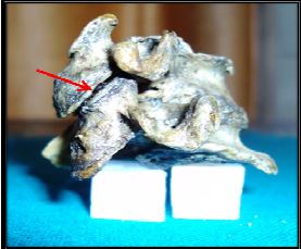
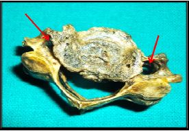
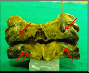
Normal typical cervical vertebrae are characterized by the presence of smaller body, foramina transversarium, triangular spinal canal. The superior articular facet are directed in upward & backward direction while the, inferior articular facet are directed forward & downwards and has a short bifid spine. Abnormalities in any of the features may be associated with neurological signs and symptoms. During the routine osteology classes we found two abnormally fused typical cervical vertebrae in the Department of anatomy, BRD medical college, Gorakhpur. These abnormalities may lead to significant sign and symptoms thus this case is being studied

Keywords: Body, Extra Foramina, Facets, Fused Cervical Vertebrae

Corresponding Author How to Cite this Article To Browse
Shalini Gupta, , Department Of Anatomy, BRD Medical college, Gorakhpur, Uttar Pradesh, India.
Email:
Bindu Singh, Prateek Gautam, Shalini Gupta, Cervical Vertebrae Synostosis & Its Clinical Significance. Biomed Rev J Basic Appl Med Sci. 2014;1(2):49-52.
Available From
https://www.biomedicalreview.in/cervical-vertebre-synostosis-its-clinical-significance-case-report

Manuscript Received Review Round 1 Review Round 2 Review Round 3 Accepted
2014-08-26 2014-08-28 2014-09-04 2014-09-11 2014-09-18
Conflict of Interest Funding Ethical Approval Plagiarism X-checker Note
Nil Nil Yes 19%

© 2014by Bindu Singh, Prateek Gautam, Shalini Guptaand Published by Siddharth Health Research and Social Welfare Society. This is an Open Access article licensed under a Creative Commons Attribution 4.0 International License https://creativecommons.org/licenses/by/4.0/ unported [CC BY 4.0].

Introduction

Cervical vertebrae are seven in number. C3-6 are typical whereas C1 (atlas), C2 (axis) and C7 (cervicae prominens) are atypical. For many years anomalies of cervical region were of interest to anatomists [1]. Congenital anomalies at cranio-vertebral or cervical region are common [2]. Among various congenital anomalies that are existing the important ones are the fused cervical vertebrae (FCV). In condition of fusion of the cervical vertebrae (FCV), two vertebrae appear structurally & functionally as one but [3]. This fusion may be congenital (CFCV) or acquired [4]. Any of the features mentioned above if found abnormal may be associated with neurological signs and symptoms having clinical importance. Since this anomaly has a clinical importance, purpose of this study is to emphasize importance of the multidisciplinary approach to help establish the precise occurrence of this anomaly for preventing any serious damage such as osteoarthritis, Klippel-Feil [5] limitation of the neck movement [6] by early treatment and diagnosis.

Case Report

During our routine osteology classes we found an abnormally fused cervical vertebra. Out of 150 typical cervical vertebrae present in the department of anatomy only one vertebra was found to be fused. The description of the fusion is under following headings:

  1. Body: Body of both the cervical vertebrae were completely fused. Lower border of the lower vertebrae was found predominantly protruded forward & downward. [Fig:-1]
  2. Articular Facets: Inferior articular facet of upper cervical vertebrae was found fused with the superior articular facet of lower cervical vertebrae on both the sides. [Fig:-2]
  3. Lamina: Unfused on both the sides.
  4. Pedicles: Unfused on both the sides.
  5. Foramen Transversarium: Present on both the sides. Foramen transversarium of left side was found to be smaller (due to osteophyte deposition) when compared from right side & also it was not aligned in same plane. [Fig:-3]
  6. Transverse Process: Unfused
  7. Coastal Elements: Unfused
  1. An extra foramen was present on the posterior lamina of upper cervical vertebrae near the inferior articular facet on right side. [Fig 4]
  2. When compared observationally for the features of this vertebrae for right & left ,it was found that the features on the right side like size of articular facet, foramen transversarium were larger compared to left side.

bm_8_01.JPG
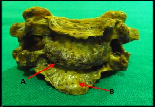
Fig 1: Arrows [A] showing complete fusion of body & [B] protrusion of lower border of lower cervical vertebraebm_8_02.JPGFig 2: Arrow showing fusion of articular facetsbm_8_03.JPG Fig 3: Arrows showing foramen transversarium of both the sides (Note the difference in size of foramen)


bm_8_04.JPGFig 4: Arrows showing presence of multiple foramen on posterior lamina of both the vertebrae.

Discussion

Fused cervical vertebrae (FCV) have both embryological as well as clinical importance. The cervical region anomalies are of interest to all like anatomists, neurologists, orthopedists, neurosurgeons. Fused vertebrae are one where two vertebra appear both structurally and functionally as one. It can be congenital or acquired due to diseases like trauma, TB, Juvenile rheumatoid arthritis, trauma or it is associated with Klippel-Feil syndrome [7].

It is important to identify the cause of FCV – whether it is congenital or acquired or pathologic condition. Clinical symptoms may vary from asymptomatic to muscular weakness, atrophy, myelopathy, limitation of the neck movement and leading to signs of peripheral nerve irritation such as,pain, burning sensations and signs of nerve compression such as hypoaesthesia/ anaesthesia, or associated with Klippel-Feil syndrome [7]. Congenital FCV is one of primary malformations of chorda dorsalis [8, 9].

Patients are generally asymptomatic, but with increasing age and injury may be symptomatic. Premature degenerative changes at adjoining motion segments are common as this condition results in greater biomechanical stress in the adjacent segments. Rupture of the transverse ligament, discal tear, spondylosis & fracture of the odontoid process are common [10].

The orthodontist may be the first person to detect cervical spine abnormalities

as they are asymptomatic until adolescence or young adulthood. Early diagnosis is based on accidental radiographic findings. There is increase in the incidence of formation of osteophyte in the adjacent levels in cervical fusion [11].

The monitorization & awareness of complications like anesthetic risk during intubation with neck extension needs to be considered.

Embryological Origin:  For the development of a normal vertebral column it is very important that the sclerotomes develops normally. But in few cases due to less blood supply or combination of genetic & environmental factors, [12] results in segmental abnormality and formation of congenitally fused vertebrae or block vertebrae.

Vertebral fusion anomalies are likely to be associated with disturbance of Pax-1 gene expression in the developing vertebral column [13].

Cervical region is the most common place for repoting this congenital fusion of vertebra, although it may involve other segments of vertebral column as well.

The location of fused vertebra in order of frequency is cervical spine (C2-3, C5-6), lumbar spine (L4-5) and thoracic spine (any section) [14)

Clinical Significance & Conclusion

In our case the excessive protrusion of lower border of lower cervical vertebrae may compress the structures present in front of it especially during hyperextension of neck. Small size of foramen transversarium & also its non-alignment on left side may cause vertebral artery compression leading to vascular signs & symptoms.

In the congenitally fused vertebrae, it is usually seen that the Antero-posterior diameter of the vertebra is decreased though the fusion may appear asymptomatic but in older age it causes degenerative changes in non-segmented cervical regions and also can leads to development of hyper mobility and degenerative changes and various other consequences. Early diagnosis of these anomalies can help in progression of degenerative process by motivating the patient to change their life style which may cause risk to the spinal cord and vertebral artery.


Reference

  1. Mcrae DL, Barnum AS. Occipitalization of the atlas. Am J Roentgenol Radium Ther Nucl Med. 1953 Jul; 70(1):23-46
  2. Romanes GJ. Cunningham`s Text Book of Anatomy.12th Ed. Oxford University Press (oxford); 1981;. 90-98.
  3. Dunsker SB, Brown O, Thomson N. Craniovertebral anomalies. Clin Neurosurg. 1980; 27:430-9.
  4. Resnick, D.: Additional Congenital or heritable anomalies and syndromes. Bone and Joint Imaging. Volume No. 2, Resnick Donald, WB Saunders Company. pp 1071-1091. (1992)
  5. de Graaff R. Congenital block vertebrae C2-3 in patients with cervical myelopathy. Acta Neurochir (Wien). 1982; 61(1-3):111-26.
  6. Bharucha Ep, Dastur Hm. Craniovertebral Anomalies: a report on 40 cases. Brain. 1964 Sep; 87:469-80.
  7. Edril H, Yidz N, Cimen M; congenital fusion of cervical vertebrae and its clinical significance; J Anat Soc India 2003;52;125-127.
  8. Besnick D, Niwayama G. Diagnosis of bone and joint disorders 2nd ed. 1985. Vol no. 5. W.B. Saunders Company, 1081-83.
  9. Meschan I. Analysis of roentgen signs in general radiology. Vol no.1.W.B. Saunders Company, Philadelphia. London. 1973; P. 618-20.
  10. Soni P, Sharma V, Sengupta J. Cervical vertebrae anomalies-incidental findings on lateral cephalograms. Angle Orthod. 2008 Jan; 78(1):176-80. doi: 10.2319/091306-370.1.
  11. Veena Vidya Shankar,Roopa R Kulkarni. Block vertebrae-Fusion of axis with third cervical vertebrae- A case report. Int J of anatomical variation; 2011; 4; 15-18
  12. Usher BM1, Christensen MN. A Sequential developmental field defect of the vertebrae, ribs and sternum, in a young woman of the 12th Cenury AD. Am J Phys Anthropol. 2000 Mar; 111(3):355-67.
  13. David KM, Coop AJ, Stevens JM, Hayward RD, Crockard HA. Split cervical spinal cord with Klippel— Feil syndrome: seven cases. Journal of Neurology. 1996; 119(6): 1859-72.
  1. Soni P, Sharma V, Sengupta J. Cervical vertebrae anomalies-incidental findings on lateral cephalograms. Angle Orthod. 2008; 78: 176-180