E-ISSN:2349-3275
P-ISSN:2349-5502

Research Article

Ventilator

Biomedical Review-Journal of Basic and Applied Medical Science

2015 Volume 2 Number 2
Publisherwww.medresearch.in

A Study on risk factors and Microbial profile of Ventilator associated Pneumonia in Intensive care units of a Tertiary care centre

Shetty S.1, Kiran V.2*, Bhat R.3, Alam N.4

1 Sanmath K Shetty, Junior resident, Department of Internal medicine, KMC, Mangalore, Karnataka, India.

2* VH Kiran, Junior resident, Department of Internal medicine, KMC, Mangalore, Karnataka, India.

3 Raghavendra V Bhat, Professor and HOD, Department of Internal medicine, KMC, Mangalore, Karnataka, India.

4 Nawaz Alam, Associate Professor, Department of Internal medicine, KMC, Mangalore, Karnataka, India.

Background: Ventilator associated pneumonia is a major cause of in hospital mortality and morbidity in our ICUs and it has received more importance with the advent of drug resistance. The knowledge of local antibiotic susceptibility patterns would guide the physician in the early empirical treatment of VAP. Our aim was to know the risk factors and microbial profile of Ventilator Associated Pneumonia patients. Methodology: We randomly studied 120 patients on ventilator in the 3 ICUs of KMC, Mangalore. VAP was diagnosed according to the case definition of VAP. The risk factors were analyzed and microbiological profile of patients who developed VAP was studied. Results: OF the 120 patients, 59 developed VAP. Prolonged ventilation and reintubation were the risk factors most significantly associated with the development of VAP. Gram negative organisms (acinetobacter, pseudomonas, klebsiella) were the most commonly isolated organisms from specimens of VAP cases and there was high prevalence of multidrug resistance. Carbapenem resistant Acinetobacter baumanii was an important pathogen in the ICU of University Medical Centre Mangalore. Vancomycin was sensitive against most gram positive isolates whereas amikacin and cefaperazone sulbactum was showing acceptable sensitivity to gram negative isolates except for Acinetobacter species. Colistin may be useful as a rescue antibiotic in carbapenem resistant Acinetobacter VAP. Conclusion: Incidence of VAP is directly proportional to duration of mechanical ventilation and Reintubation is a strong risk factor for development of VAP. Therefore, administering a proper weaning protocol and titrating sedation regimens as per the need of the patients is of utmost importance

Keywords: Pneumonia, Ventilator, Intensive care units, Drug resistance

Corresponding Author How to Cite this Article To Browse
VH Kiran, Junior resident, Department of Internal medicine, KMC, Mangalore, Karnataka, India.
Email:
Sanmath K Shetty, VH Kiran, Raghavendra V Bhat, Nawaz Alam, A Study on risk factors and Microbial profile of Ventilator associated Pneumonia in Intensive care units of a Tertiary care centre. Biomed Rev J Basic Appl Med Sci. 2015;2(2):53-58.
Available From
https://www.biomedicalreview.in/risk-factors-microbial-profile-ventilator-associated-pneumoni-intensive-care-units-research-article

Manuscript Received Review Round 1 Review Round 2 Review Round 3 Accepted
2015-07-21 2015-07-23 2015-07-30 2015-08-06 2015-08-13
Conflict of Interest Funding Ethical Approval Plagiarism X-checker Note
Nil Nil Yes 16%

© 2015by Sanmath K Shetty, VH Kiran, Raghavendra V Bhat, Nawaz Alamand Published by Siddharth Health Research and Social Welfare Society. This is an Open Access article licensed under a Creative Commons Attribution 4.0 International License https://creativecommons.org/licenses/by/4.0/ unported [CC BY 4.0].

Introduction

Ventilator associated pneumonia is a common cause of morbidity and mortality in patients admitted to an ICU. It not only has an adverse effect on patient outcome, but it also has an enormous financial impact on the patient by increasing the number of days of ventilatory support requirement, increasing cost of treatment, and also increasing the duration of stay in the intensive care unit. The incidence of VAP varies significantly across different studies and differences in VAP incidences have been based on the antibiotic profile, ICU environment, and the population of study [1]. It ranges from 6 to 52% and can reach 76% in some specific settings [2]. With the emergence of multi drug resistant “superbugs”, there is an ever increasing need for ICUs to develop an antibiotic strategy based on local epidemiological and microbiological data. Outcomes of critical patients in the ICU is to a large extent dependent on the appropriateness of the empirical antibiotic administered, which again can be formulated on the basis of local antibiotic susceptibilities [3]. Our study attempts to study the incidence of VAP in the ICUs in our institute and also identify the antibiogram profile of the microorganisms in these ICUs. We also wanted to analyze the various known risk factors for development of VAP in our study population.

Methods

The study was conducted from October 2011 to September 2013 in the MICUs of 3 teaching hospitals of KMC, Mangalore, namely University Medical Centre, Ambedkar Circle, Govt District Wenlock Hospital, Mangalore and KMC Hospital, Attavar. A total of 120 patients (40 from each ICU) who were kept on mechanical ventilator were randomly selected. Cases included were patients of both sexes who were kept on mechanical ventilator for more than 48 h. Patients who had community acquired/ hospital acquired pneumonia at intubation, patients who died within 48 hours of intubation, patients who developed pneumonia within 48 hours of intubation, patients who were discharged from the hospital before 48 hours, patients who were intubated in a different ICU and transferred to our ICU were excluded from the study. A questionnaire was prepar and each patient selected to be included in the study was screened and monitored according to the questionnaire.

Informed consent from patient bystanders was obtained. Data collection was done and patients were screened for VAP and monitored according to a proforma. Age, Sex, Date of admission to ICU, date of initiating mechanical ventilation was recorded. Patient’s vitals, general and physical examination, oxygen saturation and position of the patient were monitored regularly. Blood was sent for complete Hemogram regularly. Tracheal aspirate for culture/sensitivity was sent in patients with suspected VAP. Patients were monitored from the date of inclusion in the study to the final outcome in the ICU.

Cases of VAP was diagnosed using the case definition of VAP which is presence of a New or Progressive consolidation on chest radiology 48 hours after intubation with 2 of the following 3 features: Fever > 38° Celsius leucocytosis (>11000/mm3) or leucopenia (<4000/mm3) and purulent secretions Plus Significant growth on culture of tracheal aspirate (>105cfu/ml – quantitative or moderate to heavy growth-semiquantitative) [4].

The cases were further divided into 2 groups, “Early- onset VAP” (onset after 48 hrs but within 96 hrs) and “Late onset VAP” (onset after 96 hrs) [5]. All the patients were screened for presence of risk factors for development of VAP and occurrence of VAP in patients with the risk factors were analyzed. The culture reports were reviewed for sensitivity to the empirically started antibiotics. B on the reports of the antibiograms of the microorganisms in the 3 ICUs, an attempt was made to suggest an antibiotic policy for each of the ICUs. 

Statistical Analysis: Risk factors were assessed by deriving the relative risks for the individual risk factors. Statistical analysis for social sciences version 17.0 was used for analysis. Analysis was done by using proportions and the association estimated by chi square test. A p value less than 0.05 was considered as statistically significant.

Results

This prospective study was conducted in the 3 ICUs of hospitals of KMC, Mangalore. A total of 120 patients were studied with 40 patients from each of the 3 ICUs. The study population had an overall male predominance (64%) with an average age of 51.96 years. Among the 3 ICUs, patients in


Government wenlock hospital (GWH) ICU were younger (average age= 41.6 years) compared to University medical centre (UMC) (average age= 59.1 years) and KMC Hospital, Attavar) (average age=55.2 years).

The majority of the study population was in the 41-60 years group (33%) followed by 61- 80 years (29%) and 21-40 years (27%).There were high number of patients with infectious diseases (31), neurological problems like stroke (26), COPD(23) and poisonings(17) among the 120 cases studied. Among the 120 cases, 59 developed VAP (49%). 18 were early VAP whereas remaining 41 were late VAP cases.

The case distribution among the 3 ICUs were as shown in fig 1

bm_11_01.JPG

Fig 1: Early onset VAP and Late onset VAP

bm_11_02.JPG

Fig 2: Risk factor assessment for VAP cases

Among the risk factors studied, prolonged ventilation (> 10 days) and reintubation had the highest association with development of VAP. Male sex, unconsciousness,  immunosuppression, prior lung disease and smoking were all statistically significant risk factors for VAP.

Table 1: Risk factors associated with VAP

Reintubation Reintubation No of cases No of vapcases Percentage P value
YES 33 28 84.84% P<0.001
  NO 87 31 39.08%  
  TOTAL 120 59    
Unconsciousness Level ofconsciousness        
Conscious anddrowsy 57 20 35.08% P=0.003
  Stupor andcomatose 63 39 61.90%  
  Total 120 59    
Immunocompromised Immune status        
  Competent 73 30 41.09% P=0.025
  Compromised 47 29 61.70%  
  Total 120 59    
Lung disease lung disease        
  Present 31 21 67.74% P=0.012
  Absent 89 38 42.69%  
  Total 120 59    
Smoking Smoking        
  Smoker 27 18 66.67%% P=0.035
  Non smoker 93 41 44.08%  
  Total 120 59    
Prolonged ventilation No of days ofventilation        
  <10 days 89 31 34.83% P<0.001
  >10 days 31 28 90.32%  
  Total 120 59    

Table 2: Distribution of patients

Gender No of cases No of vap cases Percentage P value
Male 77 44 57.14% P=0.022
Female 43 15 34.88%  
Total 120 59    

bm_11_03.JPG
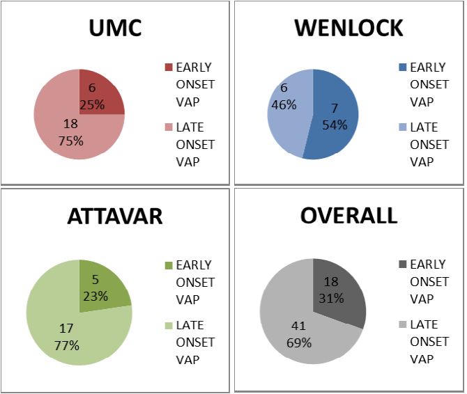
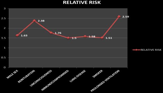
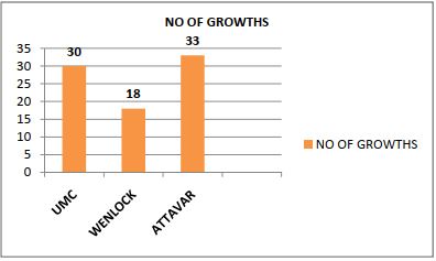
Fig 3: Isolates from 3 ICUs


A total of 81 isolates were grown from the 59 cases of VAP. The commonest organisms isolated in our study were gram negative organisms (Acinetobacter, Pseudomonas, Klebsiella) which accounted for 70.37% of all isolates followed by gram positive organisms (20.98%) and fungi (8.64%).

Table 3: Organism responsible for VAP 

UMC    
Organisms Umc Organisms(30)  
Staphylococcus Aureus 3 10%
Pseudomonas Aeruginosa 4 13.34%
Klebsiella Pneumoniae 3 10%
Acinetobacter Baumanii 14 46.67%
Enterococcus Faecalis 3 10%
Streptococcus Pneumoniae 0 0%
Citrobacter 1 3.34%
Escherichia Coli 0 0%
Proteus mirabilis 0 0%
Candida species 2 6.67%
GWH    
Organisms Wenlock Organisms (18)  
Staphylococcus Aureus 5 27.78%
Pseudomonas Aeruginosa 4 22.23%
Klebsiella Pneumoniae 3 16.67%
Acinetobacter Baumanii 3 16.67%
Enterococcus Faecalis 0 0%
Streptococcus Pneumoniae 0 0%
Citrobacter 0 0%
Escherichia Coli 2 11.11%
Proteus Mirabilis 0 0%
Candida Species 1 5.55%
KMC,Attavar    
Organisms Attavar Organisms (33)  
Staphylococcus Aureus 2 6.06%
Pseudomonas Aeruginosa 8 24.24%
Klebsiella Pneumoniae 3 9.09%
Acinetobacter Baumanii 9 27.27%
Enterococcus Faecalis 2 6.06%
Streptococcus Pneumoniae 2 6.06%
Citrobacter 0  
Escherichia Coli 2 6.06%
Proteus Mirabilis 1 3.03%
Candida Species 4 12.12%

Acinetobacter BauminiI is the commonest isolated organism in UMC and KMC Attavar and Staphyloccal Aureus is the commonest organism isolated in GWH as shown in table 3.

Table 4: Gram positive organism’s suspetibility patterns in different hospitals

Antibiotics UMC GWH KMC ATTAVAR
Amoxycillin 0 0 1
Ampicillin 0 0 2
Amoxy-clav 1 0 2
Cefaperazone 1 0 1
Clindamycin 1 3 0
Chloramphenicol 3 0 3
Erythromycin 1 1 0
Methicillin 1 0 0
Linezolid 3 2 0
Vancomycin 6 4 6
Rifampicin 3 5 2

Among the gram positive organisms, there were high degrees of resistance to commonly used antibiotics like amoxicillin, ampicillin and erythromycin in all the 3 ICUs. Vancomycin resistant strains of Staphylococcus aureus were rarely isolated (only 1), hence Vancomycin seems to the best option for empirical treatment of gram positive VAP in our ICUs.

Table 5: Gram negative organism’s susceptibility pattern

Antibiotics UMC GWH KMC
Amikacin 9 7 10
Amoxicillin 1 0 1
Ampicillin 0 0 1
Amoxy-clav 2 0 1
Ceftriaxone 3 0 1
Cefaperazone-sulbactum 11 7 18
Cefotaxime 3 1 1
Ceftazidime 5 4 2
Ciprofloxacin 4 7 4
Cotrimoxazole 1 1 4
Gentamicin 5 5 7
Imipenem 14 11 19
Meropenem 7 9 12
Piperacillin+tazobactum 5 6 7
Colistin 20    
Polymixin-B 20    
Tigecycline 6    

Among gram negative organisms, very high degrees of resistance were prevalent in our ICUs. Among the routinely usedantibiotics, amikacin sensitivity was seen in 45.61% and Cefaperazone-sulbactum sensitivity was seen in 63.15% and both these drugs may be employed for gram negative VAP


in our ICUs. Majority of the strains were resistant to other thirdgeneration cephalosporins like cefotaxime, ceftriaxone and ceftazidime and the flouroquinolones.

Discussion

The study conducted was a prospective study of 120 patients who were put on mechanical ventilation in the 3 ICUs of KMC, Mangalore. Focus was on the recognition of development of ventilator associated pneumonia in this population. 59 out of the 120 patients (49.16%) studied were diagnosed to have VAP using the case definition of VAP which was slightly higher compared to that seen in other studies [6, 7]. The high proportion of VAP cases may be attributed to the high number of stroke cases and COPD cases in our study both of which can predispose to VAP. Significant number of organophosphorus poisoning cases which leads to prolonged ventilation may also have lead to higher proportion of cases.

Male sex is associated with higher incidence of VAP. In our study, male sex was found to have a relative risk of 1.63. VAP occurred in 57.14% of males whereas it was there in only 34.88% of females. The difference was statistically significant with a p value=0.022.

Reintubation resulted in a very high incidence of VAP and proved to be an independent risk factor in various studies [8]. Reintubation as one of the risk factor for VAP this match with the study done by Hina et al [9]. This may be due to impaired reflexes after prolonged intubation or due to the altered level of consciousness, increasing the risk of aspiration. In our study 28 of the 33 cases that had to be reintubated developed VAP (84.84%). VAP incidence was 39.08% in those patients who did not require reintubation.

Immunocompromised patients like those with long standing uncontrolled diabetes, chronic kidney disease, cirrhosis of liver, HIV/AIDS, patients with blood malignancies, solid organ malignancies on chemotherapy and those on long term corticosteroids are more prone for infections including VAP. Even in our study this pattern was seen. VAP was seen in 61.70% of immunocompromised patients and only 41.09 % of immunocompetent individuals (p=0.025%).

Patients with preexisting structural lung disease

have increased incidence of VAP due to impaired defense mechanisms of the lung microstructure to microbial invasion. In this study, VAP was more prevalent in those patients with preexisting lung disease (67.74%) than in those without any lung disease (42.69%).

In our study, smoking was also found to be a risk factor; 66.67% of smokers developed VAP whereas only 44.08% of nonsmokers developed VAP (p=0.035).

In our study it was observed that incidence of Late onset VAP was more than early onset VAP this correlates with the study done by Vasudev et al [10].

The more the number of days the patient is on a ventilator, the higher is his chances of getting pneumonia. This has been proved beyond doubt in our study because 90.32% of patients who were intubated for 10 days or more had VAP whereas 34.83% of patients who were intubated for less than 10 days (p<0.001). Prolonged intubation has therefore been established as an independent and the most important risk factor for VAP in this study. Hence, it is very important for all intensivists to assess ventilated patients for weanability on a day to day basis. Superbugs are on the rise in most ICU setups [11]. In our study we have isolated MDR pathogens from VAP cases in all the 3 ICUs.

Carbapenem resistance is also very prevalent, especially in UMC ICU. The reason for this may be attributed to the rise of Acinetobacter species in this ICU. The commonly used antibiotics proved to be resistant to Acinetobacter species; amikacin was sensitive in 14.2%, amoxicillinclavulanate was sensitive in 7.14%, cefotaxime was sensitive in 7.14 %, ceftazidime was sensitive in 7.14% and ciprofloxacin was sensitive in 0%. Resistance was present to even the higher antibiotics: of the 14 growths, 8 growths were resistant to cefaperazonesulbactum, 13 were resistant to piperacillin-tazobactum, 7 were resistant to imipenem-cilastatin and 13 were resistant to Meropenem. Colistin and polymyin was sensitive in all the strains of acinetobacter. Carbapenem resistance in this ICU may be related to the increased use of carbapenems, especially meropenem in treating ICU infections over the past few years. Considering the high degree of resistance to higher antibiotics among acinetobacter


species, colistin remains the most effective antibiotic for treatment of acinetobacter VAP. However its use should be judicious in order to prevent development of resistance to this antibiotic.

Conclusion

Late onset VAP was more common than early onset VAP and was associated with multi drug resistant organisms. Male sex was an independent risk factor for development of VAP. Incidence of VAP is directly proportional to duration of mechanical ventilation and re-intubation is a strong risk factor for development of VAP. Therefore, administering a proper weaning protocol and titrating sedation regimens as per the need of the patients is of utmost importance. Unconsciousness, preexisting lung disease, smoking and immunocompromised state are all risk factors for development of VAP.

Reference

  1. Niederman MS, Craven DE. Guidelines for the management of adults with hospital-acquired, ventilator associated and healthcare-associated pneumonia. Am J Respir Crit Care Med. 2005 Feb 15; 171(4):388-416.
  2. Koenig SM, Truwit JD. Ventilator-associated pneumonia: diagnosis, treatment, and prevention. Clin Microbiol Rev. 2006 Oct; 19(4):637-57.
  3. Ulldemolins et al. Appropriateness is Critical. Crit Care Clin 2011Jan; 27(1):35–51.
  4. Davis KA. Ventilator-associated pneumonia: a review. J Intensive Care Med. 2006 Jul-Aug; 21(4):211-26.
  5. Morehead RS, Pinto SJ. Ventilator-associated pneumonia. Arch Intern Med. 2000 Jul 10; 160(13):1926-36.
  6. Arindam Dey, Indira Bairy. Incidence of multidrugresistant organisms causing Ventilator associated pneumonia in a tertiary care hospital: A nine months’ prospective study. Ann Thorac Med 2007 AprJun; 2(2):52-7.
  7. Jakribettu RP, Boloor R. Characterisation of aerobic bacteria isolated from Endotracheal aspirate in adult patients with suspected ventilator associated pneumonia in a tertiary care centre in Mangalore. Saudi J Anaesth 2012 Apr; 6(2):115-9.
  1. Torres A, Gatell JM, Aznar E, el-Ebiary M, Puig de la Bellacasa J, Gonzalez J, et al. Re-intubation increases the risk of nosocomial pneumonia in patients needing mechanical ventilation. Am J RespirCrit Care Med 1995 Jul; 152(1):137–41.
  2. Hina g, arun vyas, akshay k kar. A study of ventilatorassociated pneumonia: incidence, outcome, risk factors and measures to be taken for prevention. Indian J Anaesth. 2010 Nov-Dec; 54(6):535-40.
  3. Vasudev G, Chiranjay Mukhopadhyay, Kavitha S, PreethI V, Rishikesh K,Anathakrishna B ShastrY. Determinants of Ventilator Associated Pneumonia and its impact on prognosis: A tertiary care experience.Indian J Crit Care Med. 2013 Nov-Dec; 17(6)337-42.
  4. Joseph NM, Sistla S, Dutta T K, Badhe A S, Rasitha D, Parija S C.Ventilator-Associated Pneumonia in a tertiary care hospital in India: role of multi-drug resistant pathogens. J Infect Dev Ctries 2010; 4(4):218-225.