E-ISSN:2349-3275
P-ISSN:2349-5502

Research Article

Intracerebral Hemorrhage

Biomedical Review-Journal of Basic and Applied Medical Science

2015 Volume 2 Number 3
Publisherwww.medresearch.in

Incidence of spontaneous Intracranial Hemorrhage( ICH) & utility of the ICH scores in stroke patients

Grace M.1*, Jacob K.2, Shameer V.3, Sudhiraj.4, Vijayan S.5, Das R.6, Sasindran S.7

1* Mary Grace, Associate Professor, Department of Medicine, Government Medical College, Thrissur, Kerala, India.

2 K J Jacob, Additional Professor, Department of O & G, Government Medical College, Thrissur, Kerala, India.

3 V K Shameer, Assistant Professor, Department of Medicine, Government Medical College, Kozhikode, Kerala, India.

4 Sudhiraj, Assistant Professor, Department of Community Medicine, Government Medical College, Thrissur, Kerala, India.

5 Sarang Vijayan, Senior Resident, Department of Medicine, Government Medical College, Thrissur, Kerala, India.

6 Remya Das, Senior Resident, Department of Medicine, Government Medical College, Thrissur, Kerala, India.

7 Sooraj Sasindran, Junior Resident, Department of Medicine, Government Medical College, Thrissur, Kerala, India.

Introduction: The aim of the study was to find out the incidence of spontaneous ICH in patients hospitalized with stroke, to study the predictive value of clinical and radiological findings in determining the mortality and short term outcome and to study the utility of the ICH scores. Materials and Methods: Among the 348 consecutive patients admitted with stroke, those who were diagnosed to have primary ICH, were included in the study. The mortality and outcome were predicted using the ICH scores. Data was analyzed using SPSS software and epi info. Results: Of the 130 cases with hemorrhage there were 102 patients (29.31%) with spontaneous ICH. The cut off value of the intracerebral bleed in relation to mortality was calculated as 22.5 ml. At the end of three months 100 people were available for follow up, only 9 (21.9%) were physically dependant on others for their living. All the three ICH scores were found to be statistically significant in predicting mortality at one month, but they did not fare well in predicting the short term outcome. Conclusion: We have observed an increased incidence of hemorrhagic stroke among hospitalized patients with stroke,(29%) when compared to western studies. Volume of the hemorrhage more than 22.5 ml was associated with poor prognosis .We observed a significant improvement in the functional status at three months, with 31% of the survivors able to lead independent lives. All the three ICH scores did not fare well in predicting the short term outcome.

Keywords: Intracerebral Hemorrhage, ICH score, Mortality, Short Term Outcome

Corresponding Author How to Cite this Article To Browse
Mary Grace, Associate Professor, Department of Medicine, Government Medical College, Thrissur, Kerala, India.
Email:
Mary Grace, K J Jacob, V K Shameer, Sudhiraj, Sarang Vijayan, Remya Das, Sooraj Sasindran, Incidence of spontaneous Intracranial Hemorrhage( ICH) & utility of the ICH scores in stroke patients. Biomed Rev J Basic Appl Med Sci. 2015;2(3):68-74.
Available From
https://www.biomedicalreview.in/incidence-spontaneous-intracranial-hemorrhage-ich-utility-ich-scores-stroke-research-article

Manuscript Received Review Round 1 Review Round 2 Review Round 3 Accepted
2015-09-10 2015-09-12 2015-09-19 2015-09-26 2015-10-03
Conflict of Interest Funding Ethical Approval Plagiarism X-checker Note
Nil Nil Yes 16%

© 2015by Mary Grace, K J Jacob, V K Shameer, Sudhiraj, Sarang Vijayan, Remya Das, Sooraj Sasindranand Published by Siddharth Health Research and Social Welfare Society. This is an Open Access article licensed under a Creative Commons Attribution 4.0 International License https://creativecommons.org/licenses/by/4.0/ unported [CC BY 4.0].

Introduction

Spontaneous intracerebral hemorrhage constitutes about 10-15% of the total cases of stroke worldwide [1]. The incidence of ICH in Asian countries varies from 17-33% [2]. The overall mortality from primary ICH is approximately 40-50% [3]. Almost 40% of patients die before the 30th day after ICH, 66%of survivors are severely disabled and only 20% recover functional independence on the 6th month after ICH [4]. The aim of our study was to find out the incidence of spontaneous ICH (sICH) in patients hospitalized with stroke, to study the predictive value of clinical and radiological findings in determining the mortality and short-term outcome at three months after sICH and to study the effectiveness of the ICH scores in predicting the mortality and short term outcome. To our knowledge there are no previous similar studies on sICH done in Kerala (state in southern part of India) and there have been only very few prospective studies on ICH in India.

Materials and Methods

The cases were detected prospectively in the general medicine wards of Government Medical College Thrissur. The study period was for 7 months. Among the 348 consecutive patients admitted with clinical and radiological features of stroke, those aged more than 18 years, who were diagnosed to have primary ICH, were included in the study. The exclusion criteria were those cases of ICH secondary to trauma, bleeding into tumor, hemorrhagic transformation of infarct, subarachnoid hemorrhage, rupture of ateriovenous malformation, aneurysmal bleed, subdural hematoma, and use of oral anticoagulants. Data was collected regarding the risk factors, clinical features, laboratory investigations and neuroimaging findings. Hypertension was defined as either history of hypertension on antihypertensive drugs or recorded blood pressure more than 140/90 mm of Hg. History of diabetes, on treatment for diabetes, fasting blood sugar value more than 126 mg % or random blood sugar value more than 200 mg % was taken to be diagnostic of diabetes. Neurological assessment was done using both GCS and NIH Stroke Score [4]. The neruoimaging data collected included location of the hematoma, size of the bleed, presence of intraventricular extension and subarachnoid extension. The mortality and outcome

were predicted using the ICH scores [6]. Patients were followed up for three months from the onset of the event, to assess the short term outcome. The neurological outcome was assessed at one month and three months either by follow up in the OPD or by telephone interviews with the patient or bystander. Modified Rankin Scale (mRS) was used to assess the neurological outcome. mRS grade equal to or less than 2 was taken as good outcome and values equal to or more than three was regarded as poor outcome. [7].

Statistical analysis: Data was analysed using SPSS software version 16 and epi info. All continuous variables were analyzed with unpaired t test for normally distributed data and Mann-Whitney U test for skewed data. Categorical variables were analyzed using Chisquare test. We also derived cutoff values using ROC curve with optimal sensitivity and specificity for, volume of hematoma, GCS and NIHS scores.

Results

Among the 348 patients admitted with cerebrovascular disease 102 patients with spontaneous intracerebral hemorrhage were included in the study as shown in Figure 1

bm_14_01.JPGFigure 1

The mean age of the patients with spontaneous ICH was 63.7 ±14years. The mean age of males was 61.9±14 years and that of females was 67.5 ±13years. There were 69 males (67.65%) and 33(32.35%) females. In the study cohort of 102 patients, hypertension was seen in 90.2%, diabetes in 26%, alcohol consumption in 26%, smokers 30% and history of coronary artery disease 6.9%. The commonest mode of presentation was altered level of consciousness. Mean systolic blood pressure


at the time of presentation was 177±36 mm of Hg and mean diastolic blood pressure was 100±17.5 mm of Hg. Median±IQR GCS score on admission was 8.5±11 and Median±IQR NIHS score was 17±13.

The locations of the IC bleed and their frequencies were as follows capsuloganglionic 50 (49.02%), lobar 29 (28.43%), cerebellum 8 (7.8%), thalamic 4 (3.92%), brainstem and intraventricular hemorrhage 3 each (2.94%) and 5 patients had bleeding in multiple locations (5%). Intraventricular extension of the bleed was seen in 55(53.92%). There was statistically significant correlation between neurological assessment scores and hematoma volume (NIHS score Spearman’s rho-.412 p value <.001, GCS score Spearman’s rho -.241 p value .015). There was also statistically significant negative correlation between blood sugar level and the GCS score (Spearman‘s rho -.301 p value .008). Blood sugar level was significantly associated with volume of bleed (Spearman’s rho-.235 p 0.019.)

Mortality at one month: The association of demographic and clinical profile of the patients is shown in table 1.

Table 1: Association of demographic and clinical profile of ICH with one month mortality

Variables Aliven (%) Deadn (%) Risk ratio Lowerlimit Upperlimit
GenderMaleFemale 25(36.7%)16(50%) 43(63.2%)16(50%) 1.26 .85 1.86
diabetes 8 (29.6%) 19(70.3%) 1.2 .9 1.7
hypertension 38 (41.7%) 53(58.2%) .87 .53 1.4
History of ischemic heartdisease 3(42.8%) 4(57%) .9 .49 1.8
headache 10 (52%) 9 (47%) .767 .463 1.27
Altered level ofconsciousness 14(22.5%) 48(77.4%) 2.67 1.59 4.48
Systolic BP >140mm ofHg 36(40.9%) 52(59%) 1.01 .609 1.68
GCS score3,45-1213-15 1(3.57%)13(33.3%)27(81%) 27(96.43%)26(66.67%)6(18%) 2.13.66 1.61.7 2.87.8
NIHS score>2011-200-10 717%)18(51%)16(64%) 34(83%)17(50%)8(33%) 1.91.45 1.4.75 2.72.8
Volume of hematoma<30ml30ml-60ml>60ml 31(52%)9(37.5%)1(7.14%) 29(48%)15(62.5%)13(92.86%) 1.21.92 .861.42 1.92.59
Intraventricular extension 7(13.2%) 46(86.7%) 3.1 1.9 5

At the end of one month, out of the 100 patients with intracerebral bleed, 57 had died. The mean age of those who survived was 62.1±14.2 years and of those who died was 64.1 ± 13.7 years. There was no statistically significant difference in the mortality at one month with respect to age, sex, presence of co morbidities like diabetes, hypertension, and previous history of coronary artery disease or cerebrovascular disease. Headache, vomiting, seizures or the presence of weakness at the time of presentation did not affect the mortality. Alteration in the level of consciousness carried an increased risk of mortality. Blood pressure at the time of admission did not show any relation to mortality. Location of the bleed and subarachnoid extension of the hemorrhage did not influence the mortality.

The factors which were found to have statistical significance in predicting mortality were altered level of consciousness on presentation, assessment of neurological status by GCS and NIHS scores, volume of bleed, intraventricular extension of bleed. Multiple regression analysis showed that only GCS score, volume of bleed and intraventricular extension showed statistical significance (pvalue <.001 Cox and Snell .461).

The cut off value of the intracerebral bleed in relation to mortality was calculated as 22.5 ml [70% sensitivity and 68% specificity AUC .716(.614-.818)], for the GCS score the cut off value was 7 [sensitivity 66%, specificity90% AUC .851(.775-.926)], for the NIHS score the cut off value was 15, [73% sensitivity, 787% specificity AUC .754(.653-.854)] above which the prognosis was poor.

Short term outcome: The short term outcome is shown in figure 2

bm_14_02.JPGFigure 2


*2 people were lost to follow up

The association of parameters with the short term outcome is depicted in table 2.

Table 2 Association of demographic and clinical profile of patients with ICH with their outcome at 3rd month

Variables Bad outcome Good outcome Risk ratio Upper Lower
Gendermalefemale 4(14.8%)5(35.7%) 23(85.2%)9(64.3%) .4152.4 .132.76 1.37.5
HT 9(24.3%) 28(75.6%)      
History of ischemic heartdisease 2(66.7%) 1(33.3%) 3.6 1.2 10
Diabetes 2(66.7%) 1(33.3%) 3.3 1.1 9.5
GCS score5-1213-15 5(58%)4(13.7%) 7(42%)25(86.2%) 3 .97 9.3
NIHS score0-10>20 2(11.1%)3(60%) 16(88.89%)2(40%) 5.4 1.21 23.9

Of the 43 patients who were surviving at the end of one month physically dependant on others for their living.

At the end of three months 41 were surviving, 32(78%) were able to lead independent lives while only 9(21.9%) were physically dependant on others for their living.

The mean age of those living unaided was 62.8± 13.9 years and that of those physically dependant was 56.5 ± 11.2 years. Females were more likely to have a bad outcome than males. Presence of hypertension was found to be a predictor of bad outcome.

The factors which had statistical significance in relation to bad outcome of patients are diabetes, history of ischemic heart disease, GCS score and NIHS score.

The factors which influenced mortality like altered level of consciousness, volume of bleed and intraventricular extension do not affect the disability. The utility of the ICH scores were compared as shown in figure 3 and 4

All the three ICH scores were found to be statistically significant in predicting mortality at one month. Utilizing the ROC the modified ICH score had the best AUC .875(.805-.944) among the three scores in predicting mortality at one month. All the three ICH scores did not fare well in predicting the short term outcome (original ICH Score .665, new ICH score .58 and modified ICH score .606)

bm_14_03.JPG
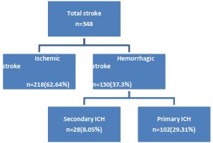
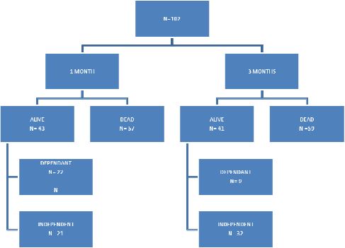
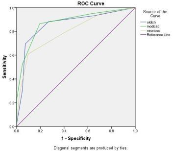
Figure 3: ROC curve comparing the ICH scores in predicting one month mortality

bm_14_04.JPG
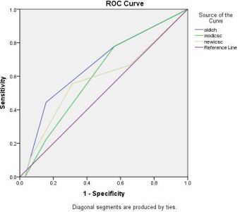
Figure 4: ROC curve of the ICH scores in predicting short term outcome

Discussion

The clinical outcome of intracerebral hemorrhage is generally poor. It is important to identify the factors which influence the mortality and prognosis of ICH.

Globally ischemic strokes account for 85% and hemorrhage account for 15% of all strokes. According to data from the Asian Stoke Advisory Panel ASAP, the incidence of ICH in Asian countries varies from 17- 33%.[2].

The percentage of ICH among stroke was found to be 55.4% in a Chinese study, [7]., 42% in Pakistan, [9]. 40%-50% in Thailand, [10] & 32-42% in India [11].


Of the 348 patients admitted with cerebrovascular disease there were 218 cases of ischemic stroke (62.64%), and 130 cases (37.3 %) of hemorrhagic stroke. Of the 130 cases with hemorrhage there were 102 patients (29.31%) with spontaneous ICH and 28 cases (8.05%) with secondary ICH. Thus we also have observed an increased incidence of hemorrhagic stroke among hospitalized patients, when compared to western studies.

The common modes of presentation in ICH are weakness, altered level of consciousness, headache vomiting and seizures. In this study altered level of consciousness was the most common way of presentation. Seizures are usually associated with lobar hemorrhage, while headache is commonly seen in cerebellar hemorrhage. [12]. we also observed that among those who had seizures at the onset 58% had lobar hemorrhage. In most of the studies, the commonest site of bleed is capsuloganglionic (71%) followed by thalamus (17%), cerebral hemispheres (4%), brainstem (7%) and cerebellum (1.4%) in the descending order of frequency. [13,14]. In our study capsuloganglionic continued to be the commonest location of ICH (49%), but there were more cases of lobar (28%) and cerebellar bleed (8%) compared to other studies. Some studies have shown that diabetics tend to have a larger volume of bleed. [15]. We also observed significant association between diabetes and ICH volume.

The overall mortality from primary ICH is approximately 40-50% [13]. Almost 40% of patients die before the 30th day after ICH .We observed a mortality rate of 57% at one month, which was much higher when compared to other studies. 30day mortality reported from other studies are Turkey 38.3% [16]. European study 37%, and study from Pakistan 39.7%.

In this study the variables which are statistically significant in predicting mortality at one month are assessment of neurological status by GCS, volume of bleed, presence of intraventricular extension of bleed. A high BP at the time of admission was found to be a factor contributing to the poor outcome in some studies [14]. We did not get any significant correlation between blood pressure and mortality. A high blood sugar value on admission has been implicated as an independent predictor of death after ICH. [14, 16]. We observed that diabetes is not a predictor of mortality at one month.

Many of the studies have observed that volume of bleed more than 30 ml (42 ml in a study from AIIMS) [15]. And presence of intraventricular extension of bleed are predictors of poor prognosis. Using the ROC we have obtained a cut off value of the intracerebral bleed of 22.5 ml (70% sensitivity and 68% specificity AUC .716) above which the prognosis is poor.

Short term outcome: Between 32 and 50% die within the first month after the event and only 20% are independent at six months after intracerebral bleed. [12]. Proportion of patients who were able to lead an independent life after one year varies between 12 and 39.35%. In our study there were only 43 patients surviving at the end of one month, and only 21% of patients were leading independent lives. But we observed a significant improvement in the functional status at three months, with 31% of the survivors able to lead independent lives.

The factors which had statistical significance in relation to bad outcome of patients are hypertension, diabetes, history of ischemic heart disease and NIHS score. There was no significant difference in the outcome at one month with respect to sex, history of previous cerebrovascular disease, mode of presentation, location of the bleed, volume of bleed, intraventricular extension and subarachnoid extension of the hemorrhage. The factors like altered level of consciousness, volume of bleed, intraventricular extension which are significant predictors of mortality at one month, have failed to achieve significance in predicting whether the person who survives the event would be able to lead independent lives.

ICH Scores: We observed that all the three ICH scores are significant in predicting mortality at one month. The advantage of the new and modified ICH score is that volume of the bleed is not included as a variable, thus obviating the fallacies which may be associated with calculation of the volume of bleed using the formula 4/3πabc. [17]. None of the three ICH scores were good enough to predict the short term functional outcome.

The limitations of our study were the small sample size, long term outcome had not been studied, sequential CT scans were not taken in the majority of cases, and it was not a population based study. Strength of our study was that all the patients were seen consistently by at least one designated


investigator and we could get the follow up at three months in the majority of the patients (only 2 patients were lost to follow up).

Conclusions

We observed an increased incidence of hemorrhagic stroke among hospitalized patients with stroke,(29%) when compared to western studies. Volume of the hemorrhage more than 22.5 ml was associated with poor prognosis 70% sensitivity and 68% specificity). The variables which were independent predictors of mortality at one month were poor neurological status as assessed by GCS and NIHS scores, larger volume of bleed, and presence of intraventricular extension of bleed. There was no statistically significant correlation between blood pressure, diabetes, location of bleed and the mortality.

We observed a significant improvement in the functional status at three months, with 31% of the survivors able to lead independent lives. This is in marked contrast to the existing data wherein only around 20% achieve functional independence by about six months.

The factors which had statistical significance in relation to bad outcome of patients are the presence of comorbidities like hypertension, diabetes, history of ischemic heart disease and the assessment of neurological status by the NIHS score. We observed that all the three ICH scores were good in predicting mortality at one month, but not so to predict the short term functional outcome.

Our study despite its limitations has succeeded in establishing the clinical and radiological parameters which have an influence on early mortality and short term outcome. This information is useful for early stratification of patients who could improve under intensive care, from those with suggested poor outcome, and for better communication with patients and their relatives.

Reference

  1. Broderick J P, Brott T, Tomsick T, Miller R HG. Intracerebral hemorrhage more than twice as common as SAH. J Neurosurg. 1993; 78(2):188–91.
  2. Asian Acute Stroke Advisory Panel (AASAP). Stroke epidemiology data of nine countries. J Med Assoc Thai. 2000; 1:1–7.
  1. S F Sia, K S Tan VW. Primary Intracerebral hemorrhage in Malaysia: In hospital mortality and Outcome in patients from a hospital based Registry. Med J Malaysia. 2007; 62(4):308–12.
  2. Linda S Williams,Engin Y Yilmaz,Alfredo M LopezYunez Retrospective assessment of initial stroke severity with the NIH Stroke Scale 200;31:858-862
  3. Ribo M GJ. Latest advances in intracerebral hemorrhage. Curr Neurol Neurosci Rep. 2006; 6(1):17–22.
  4. Cheung RT1 ZL. Use of the original, modified, or new intracerebral hemorrhage score to predict mortality and morbidity after intracerebral hemorrhage. Stroke. 2003; 34(7):1717–22.
  5. Ruijun Ji, Haipeng Shen, Yuesong Pan, Penglian Wang, Gaifen Liu YW. A novel risk score to predict 1- year functional outcome after intracerebral hemorrhage and comparison with existing scores. Crit Care. 2013; 17(6):R275.
  6. Yang QD, Niu Q, Zhou YH, Liu YH XH et al. Incidence of cerebral hemorrhage in the Changsha community. A prospective study from 1986 to 2000. Cerebrovasc Dis. 2004; 17(4):303-13
  7. Shams-ur-Rehman and Muhammad Ali Khan Khan SR and MA. CLINICAL VERSUS C.T. SCAN DIAGNOSIS IN STROKE–A COMPARATIVE STUDY OF 50 CASES. J Ayub Med Coll Abbottabad. 2002; 14(1):2–5.
  8. Daga MK, Sarin K N V. Comparison of Siriraj and Guy’s Hospital score to differentiate supratentorial ischaemic and haemorrhagic strokes in the Indian population. S. J Assoc Physicians India. 1994; 42(4):302– 3.
  9. Ashraf V Valappil, Nilesh V Chaudhary, R Praveenkumar, Biju Gopalakrishnan AG. Low cholesterol as a risk factor for primary intracerebral hemorrhage: A case-control study annals of indian academy of neurology. 2012; 15(1):19–22.
  10. Zeynab Mayabi, Mahmoud Omrani MG. Spontaneous Intracerebral Hemorrhage; Findings of Primary CT-scan, Clinical Manifestations and Possible Risk Factors. J MedSci. 2013; 13:124–9.
  11. Sunil K Narayan, P Sivaprasad,

  1. Sharma Sushma, Ratnakar K Sahoo TKD. Etiology and outcome determinants of intracerebral hemorrhage in a south Indian population, a hospital-based study. Ann Indian Acad Neurol. 2012; 15(4):263–6.
  2. Bhatia R, Singh H, Singh S, Padma M V, Prasad K, Tripathi M, Kumar G SMB. A prospective study of inhospital mortality and discharge outcome in spontaneous intracerebral hemorrhage. Neurol India. 2013; 61:244–8.
  3. Maya P. Danovska, Margarita L. Alexandrova, Nachko I. Totsev, Irena I.Gencheva PGS. Clinical and neruoimaging studies in patients with acute spontaneous intracerebral hemorrhage. J IMAB - Annu Proceeding Scientific Pap. 20(2):489–94.
  4. Börü ÜT, Gül L TM. A hospital- based study on long term mortality and predictive factorsafter spontaneous intracerebral hemorrhage from Turkey. Neurol Asia. 2009; 14:11–4.
  5. Broderick JP, Brott TG, Duldner JE, Tomsick.T HG. Volume of Intracerebral Hemorrhage: a powerful and easy to use predictor of 30-day mortality. Stroke. 1993; 24:987–93