E-ISSN:2349-3275
P-ISSN:2349-5502

Case Report

Mucocele of appendix

Biomedical Review-Journal of Basic and Applied Medical Science

2016 Volume 3 Number 1
Publisherwww.medresearch.in

Mucocele of appendix in an elderly male from tuberculosis endemic region

Khadanga S.1*, Dugar D.2, Khurana U.3, Thatoi P.4, satapathy S.5

1* Sagar Khadanga, Assistant Professor, Department of Medicine, AIIMS, Bhopal, Madhya Pradesh, India.

2 Dharmendra Dugar, Assistant Professor, Department of Surgery, Hitech Medical College, Bhubaneswar, Odisha, India.

3 Ujjawal Khurana, Assistant Professor, Department of pathology, People’s College of Medical Sciences, Bhopal, Madhya Pradesh, India.

4 Pravat Kumar Thatoi, Assistant Professor, Department of Medicine, SCB medical college, Cuttack, Odisha, India.

5 Shakti Prasad satapathy, Senior consultant, Department of Medicine, Aditya care hospital, Bhubaneswar, Odisha, India.

A 55 year old gentleman with past history of pulmonary tuberculosis 6 years back presented with diffuse abdominal pain associated with fever, vomiting and a vaguely palpable mass in right iliac fossa. Ultrasonography showed distended ileal loops in the right lower quadrant of abdomen. CT scan abdomen revealed thickened, enlarged and fluid filled appendix suggestive of mucocele. Laparoscopy revealed approximately 22cm X 5cm mucocele of appendix with omental adhesions. Laparoscopic appendectomy was done, without any spillage of mucin in the peritoneum. The patient was discharged from the hospital on 3rd postoperative day and on ten months of follow up the patient is doing well. Laparoscopic appendectomy do not increase complication rate in mucocele of appendix

Keywords: Mucocele of appendix, Mucinous cystadenoma, Pseudomyxomaperitonei

Corresponding Author How to Cite this Article To Browse
Sagar Khadanga, Assistant Professor, Department of Medicine, AIIMS, Bhopal, Madhya Pradesh, India.
Email:
Sagar Khadanga, Dharmendra Dugar, Ujjawal Khurana, Pravat Kumar Thatoi, Shakti Prasad satapathy, Mucocele of appendix in an elderly male from tuberculosis endemic region. Biomed Rev J Basic Appl Med Sci. 2016;3(1):153-155.
Available From
https://www.biomedicalreview.in/mucocele-of-appendix-in-an-elderly-male-from-tuberculosis-endemic-region-case-report

Manuscript Received Review Round 1 Review Round 2 Review Round 3 Accepted
2016-03-08 2016-03-10 2016-03-17 2016-03-24 2016-03-31
Conflict of Interest Funding Ethical Approval Plagiarism X-checker Note
Nil Nil Yes 17%

© 2016by Sagar Khadanga, Dharmendra Dugar, Ujjawal Khurana, Pravat Kumar Thatoi, Shakti Prasad satapathyand Published by Siddharth Health Research and Social Welfare Society. This is an Open Access article licensed under a Creative Commons Attribution 4.0 International License https://creativecommons.org/licenses/by/4.0/ unported [CC BY 4.0].

Introduction

Mucocele of appendix is an obstructive dilatation of appendix with intra luminal mucoid material accumulation. It is a rare lesion found in only 0.2% to 0.3% of appendectomies [1]. The non-specific pain in lower abdomen may be mistaken for typhlitis, ileo colitis or acute appendicitis. If the symptoms persist over weeks it may be confused with tuberculosis or inflammatory bowel disease. In spite of extensive investigations, the preoperative diagnosis may remain elusive. Surgical resection (appendectomy) is the method of choice in management of simple mucocele and cystadenoma with an intact base [2]. Some studies of successful laparoscopic resection of mucocele have been already reported [3].

Case Report

A 65 year old man with past history of completed antitubercular therapy for pulmonary tuberculosis 6 years back presented with complaints of fever and pain in lower abdomen for 1 month with point of maximum intensity in the right iliac fossa (RIF). The fever was irregular and low grade. During this period he was vomiting once or twice a day associated with altered bowel habits. Physical examination revealed pulse- 108/min, blood pressure 130/86mm Hg, respiratory rate-18/min and temperature-100.40F. Abdominal examination revealed muscle guarding with rebound tenderness in the RIF. There was an ill-defined soft mass of 15x8cm in the RIF. Cardiovascular, respiratory and central nervous system examination did not reveal any abnormality. Routine hemogram revealed neutrophilic-pleocytosis (14,200/cmm, 76%). Kidney and liver functions were normal. Ultrasonography showed gas filled small intestinal loops in the RIF with probe tenderness, suggesting acute appendicitis. CT scan of abdomen revealed cystic dilatation of appendix22x8cm with surrounding inflammation. A provision diagnosis of mucocele of appendix was made with other cystic appendicular masseswere kept as differentialdiagnosis (mucinous cystadenoma, mucosal hyperplasia, mucinous cystadenocarcinoma and retention cyst). We planned for a laparoscopic appendectomy. Pneumo-peritoneum was created with Veress needle using C02 and the table was kept in Trendelenburg position with 15 degree tilt to left. A 300 telescope was introduced through

umbilical port and complete examination of abdomen was done. Laparoscopy revealed approximately 25x10cm mucocele of appendix with omental adhesions. The walls were inflamed but without any perforation. No discharge was found in peritoneal cavity. The mucocele was isolated and separated with the help of bipolar cautery (Figure-1). The base was ligated, divided and the appendix was retrieved out in a plastic bag through the umbilical port. Only appendecectomy was performed as there was no pathological process at the base of appendix or mesenteric lymphadenopathy. The specimen was sent for histopathology (Figure-2).

The patient was discharged from the hospital on 3rd postoperative day. Later the histopathology confirmed the diagnosis of mucinous cystadenoma of appendix. Histopathological features showed appedicular lumen distended with mucus. The lining epithelium showed flattened epithelium with dysplastic lining. However there was no foci of invasion or dissection of mucin through the appendiceal wall (Figure-3). The patient is followed up for 10 months and was doing fine.

bm_26_01.JPG
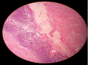
Fig 1: Intra operative image of laparoscopic appendectomy for mucocele.
bm_26_02.JPG
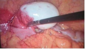
Fig-2: Cut specimen of appendix showing mucin.


bm_26_03.JPG
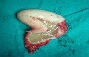
Fig 3: Histopathology showing appendix wall lined with mucinous epithelium.

Discussion

Mucocele of appendix is a term used for appendix distended by mucous secondary to mucinous cystadenoma (63%), mucosal hyperplasia (25%), mucinous cystadenocarcinoma (11%) and retention cyst (6%). Clinical presentation is nonspecific. Approximately 25-30% of patients are asymptomatic and discovered incidentally [4]. Ultrasonography is often the first line imaging modality but may not be very sensitive. Appndicular-mass can be purely cystic or complex hypoechoic masses with fine internal echoes [5].

Appendicular diameter >15mm indicates mucocele with 3% sensitivity and 92% specificity [6]. CT scan is regarded as better imaging with lumen >13mm, cystic dilatation and wall calcification. In absence of any distinct guideline appendectomy is considered appropriate treatment in unruptured benign mucocele. Local invasion and involvement of caecum are indications for right-hemicolectomy [7].

Laparoscopic appendectomy can be safely done by taking precautions by using bowel graspers and using a non- permeable bag. Postoperative follow up should be done to rule out subsequent pseudomyxoma peritonei.

Conclusion

CT imaging of appendicular mass is necessary and has an important role in selection of surgical technique to reduce the chances of pseudomyxoma peritonei.

We believe that laparascopic appendectomy do not increase complication rate if done carefully, especially while handling and removing the mucocele from the abdomen.

References

  1. Stang A, Braumann D, Teichmann W. [Mucocele of the appendix. Incidental sonographic discovery and laparoscopic resection]. Dtsch Med Wochenschr. 2004 Oct 22; 129(43):2295-8.
  2. Stocchi L, Wolff BG, Larson DR, Harrington JR. Surgical treatment of appendiceal mucocele. Arch Surg. 2003 Jun; 138(6):585-9; discussion 589-90.
  3. Lau H, Yuen WK, Loong F, Lee F. Laparoscopic resection of an appendiceal mucocele. Surg Laparosc Endosc Percutan Tech. 2002 Oct; 12(5):367-70.
  4. Soweid AM, Clarkston WK, Andrus CH, Janney CG. Diagnosis and management of appendiceal mucoceles. Dig Dis. 1998 May-Jun; 16(3):183-6.
  5. Skaane P, Ruud TE, Haffner J. Ultrasonographic features of mucocele of the appendix. J Clin Ultrasound. 1988 Oct; 16(8):584-7.
  6. Lien WC, Huang SP, Chi CL, Liu KL, Lin MT, Lai TI, Liu YP, Wang HP. Appendiceal outer diameter as an indicator for differentiating appendiceal mucocele from appendicitis. Am J Emerg Med. 2006 Nov; 24(7):801-5.
  7. Zagrodnik DF 2nd, Rose DM. Mucinous cystadenoma of the appendix: diagnosis, surgical management, and follow-up. Curr Surg. 2003 MayJun; 60(3):341-3