E-ISSN:2349-3275
P-ISSN:2349-5502

Research Article

Auditory and Visual cues

Biomedical Review-Journal of Basic and Applied Medical Science

2017 Volume 4 Number 1
Publisherwww.medresearch.in

Effectiveness of auditory and visual cues on Bradykinesia in individuals with Parkinson’s disease: a comparative study

Joshi S.1*, Deshpande A.2

1* Snehal Joshi, Professor, D.E. Society’s Brijlal Jindal College of Physiotherapy, Pune, Maharashtra, India.

2 Arohi Deshpande, Intern, D.E. Society’s Brijlal Jindal College of Physiotherapy, Pune, Maharashtra, India.

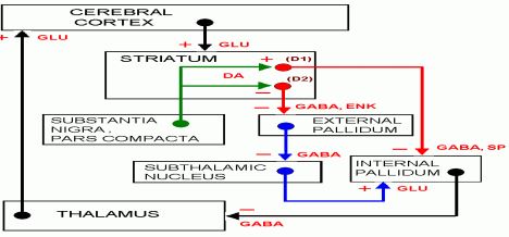
Introduction: Parkinson’s disease (PD) is a neurodegenerative disorder affecting the physical, psychological, social, and functional status of an individual. Individuals with Parkinson’s disease (PD) often demonstrate bradykinesia during mobility tasks. Due to bradykinesia; a person with Parkinson’s may have difficulty performing everyday functions. This leads to activity limitation and participation restriction in these individuals. Hence different strategies are needed to overcome bradykinesia. Use of visual cues and auditory cues can be thought of improving this problem. Hence this study was conducted to find out the efficacy of auditory cues and visual cues and compare them. Objectives 1)To assess the effectiveness of auditory cues on bradykinesia in Parkinson’s patients 2) To assess the effectiveness of visual cues on bradykinesia in Parkinson’s patients. 3) To compare the effectiveness of auditory and visual cues on bradykinesia in Parkinson’s patients. Methodology: After obtaining consent from the subjects, they were randomly divided into 3 groups in where they received either auditory cues (Metronome beats) or visual cues (video) or no cues for the selected functional activities. Reaction time was measured before and after intervention. Conclusion: It was concluded that auditory cues are effective in improving bradykinesia

Keywords: Auditory cues, Bradykinesia, Parkinson’s disease, Visual cues

Corresponding Author How to Cite this Article To Browse
Snehal Joshi, Professor, , D.E. Society’s Brijlal Jindal College of Physiotherapy, Pune, Maharashtra, India.
Email:
Snehal Joshi, Arohi Deshpande, Effectiveness of auditory and visual cues on Bradykinesia in individuals with Parkinson’s disease: a comparative study. Biomed Rev J Basic Appl Med Sci. 2017;4(1):190-196.
Available From
https://www.biomedicalreview.in/effectiveness-auditory-visual-cues-bradykinesi-individuals-parkinson-s-disease-research-article

Manuscript Received Review Round 1 Review Round 2 Review Round 3 Accepted
2017-05-08 2017-05-09 2017-05-16 2017-05-23 2017-05-30
Conflict of Interest Funding Ethical Approval Plagiarism X-checker Note
Nil Nil Yes 18%

© 2017by Snehal Joshi, Arohi Deshpandeand Published by Siddharth Health Research and Social Welfare Society. This is an Open Access article licensed under a Creative Commons Attribution 4.0 International License https://creativecommons.org/licenses/by/4.0/ unported [CC BY 4.0].

Introduction

Parkinson’s disease (PD) is a neurodegenerative disorder affecting the physical, psychological, social, and functional status of an individual [1]. It is a progressive disease associated with a degeneration of the dopamine-producing cells in the substantia nigra. In recent years, it has been highlighted that PD is a multisystem neurodegenerative disorder with motor as well as non-motor features [2]. Among motor symptoms and signs, the cardinal features (bradykinesia, rest tremor, and rigidity) are mainly due to the loss of dopaminergic neurons [3], but those involving posture, balance, and gait are largely secondary to degeneration of nondopaminergic pathways. These problems significantly contribute to impairment and disability in advanced PD patients [4]. The basal ganglia (BG) include the striatum, which comprises the caudate nucleus, putamen, and nucleus accumbens, the globus pallidus that is divided into an external segment (GPe) and an internal segment (GPi), the substantia nigra that can be divided into a pars compacta (SNc) and a pars reticulata (SNr), and the subthalamic nucleus (STN) [5]. The main input region of the BG is the striatum, which receives afferents from many regions of the cerebral cortex, including motor and premotor, cingulate, and prefrontal cortices, and the intralaminar nuclei of the thalamus [5–7].

The major output regions of the BG are the GPi and the SNr, which project to the thalamus modulating activity of cortical regions and to the brainstem [5-7]. The input and output regions are connected via either the direct or the indirect pathways, both of which arise from the matrix medium spiny neurons of the striatum, while the striosomal medium spiny neurons control dopaminergic projections from the SNc [5–8]. Corticostriatal projections, intrinsic BG circuits, and output pathways are functionally arranged according to the BG loop involved [7, 8]. The main neurotransmitter of BG circuit is the inhibitory gamma-aminobutyric acid (GABA), while neurons of the STN use excitatory glutamate and those of the SNc use dopamine [9]. According to this model, the pathophysiological cause of PD hypokinetic signs is the prevalence of the indirect pathway over the direct one resulting in increased neuronal firing activity in the output nuclei of the BG and leading to excessive inhibition of thalamocortical and brainstem motor systems

leading to alteration in normal speed of movement, onset and execution [5–6]. At variance, overactivity in the direct pathway and imbalance with the indirect one may cause reduced inhibitory BG output causing dyskinesia, PD [7, 10].

Bradykinesia refers to slowness of movement that is ongoing, akinesia indicates failure of voluntary, spontaneous (e.g., in facial expression), or associated movement (e.g., arm swing during walking) to occur, and hypokinesia refers to movements that are smaller than desired, in particular with repetitive movements [11]. In addition to whole-body slowness, bradykinesia may impair the fine motor movements, which is usually demonstrated in PD patients during rapid alternating movements of fingers, hand, or feet as a progressive reduction of speed and motion amplitude [11]. Bradykinesia is represented cranially by loss of facial expression, decreased frequency of blinking, monotonic and hypophonic speech, and drooling due to decreased spontaneous swallowing. Other manifestations of bradykinesia are slowness in rising from a chair, loss of spontaneous gesturing, reduction of handwriting (micrographia), reduced arm swing when walking, and reduced gait amplitude and velocity [11].

Although both speed and movement amplitudes are affected in PD, the speed is usually more affected, suggesting that they may be associated with partially separate mechanisms [12]. The pathophysiology of bradykinesia is not completely understood, but amongst the theories putforth, failure of the BG output to reinforce the cortical mechanisms may involve the preparation of the movement or its execution [5-7, 11]. Deficits in movement preparation in PD patients have been documented by slower reaction times [13, 14] and slower increase in premovement cortical excitability [15]. Bradykinesia combined with other PD-related movement deficits may contribute to self-reported reductions in quality of life [16]. It is one of the common reasons for hindrance in daily routine activities in these patients. This may result in deterioration of patient’s psychological and social state of mind.

Hence various interventions have been developed to improve bradykinesia. A study conducted to find out the effectiveness of high resistance training showed that Muscle force, bradykinesia, and QOL were improved to a greater degree in


those that performed high intensity eccentric resistance training compared to an active control group [17]. A Meta-analysis of effectiveness of exercise interventions for people with Parkinson’s disease reported a statistically significant benefit in exercise intervention [18]. Being debilitating, bradykinesia needs to be given attention and therefore an effective strategy should be used along with the conventional treatment so as obtain maximum benefit. Various researchers have proved that auditory and /or visual cues are effective in improving bradykinesia. A study conducted to find out the effects of pulsed auditory stimulation on various gait measurements in persons with Parkinson's disease confirmed previous findings that auditory stimulation can be used to positively influence the gait of persons with PD [19]. Another study conducted to find Effects of visual and auditory cues on gait in individuals with Parkinson's disease stated that the Visual and auditory cues improved gait performance in patients with PD. Also auditory cueing significantly improved cadence, but visual cueing improved stride length. The simultaneous use of auditory and visual cues did not improve gait significantly more than each cue alone [20]. Even though there are various studies which studied the effectiveness of cues on gait, there is dearth of studies which intended to find out the effectiveness of cues on bradykinesia. Hence this study was conducted to find out the effectiveness of cueing on bradykinesia on Parkinson’s patients and to compare the efficacy of auditory and visual cues.

Methodology

Type of study: Cross sectional study

Type of sampling: Simple Random Sampling

Number of samples: 30

Individuals suffering from Parkinson’s disease graded I&II on Hoen-Yahr scale and who were independent ambulators, were included in the study. Individuals with known visual or auditory deficits, cognitive impairments, psychiatric disorders and other neuromuscular or musculoskeletal disorder were excluded from the study.

Procedure: After obtaining approval from institutional ethics committee, consent from subjects, the subjects were randomly divided into three groups Group A, B and C. Subjects were

made to sit on chair comfortably and were asked to reach for the glass kept on the table kept around 18 inches away from the subject. Reaction time for upper limb activity that is reaching for the glass and reaction time was measured using stopwatch. 3 trials were given and best of the 3 was taken. Then the subjects were given rest period of 5 minutes rest period and were asked to do spot marching. The reaction time was measured. One group was given auditory cues in the form of metronome (Group A), one group was given visual cues in the form of video clip (Group B) and one group did not receive any cues (control Group, Group C). In auditory cues group (Group A), five times metronome beats were played and the subjects were asked to perform the activities on the pace of the beats. The second group (group B) was given visual cues in the form of video clips of the task and was asked to perform repetitions along with the video. The third group that was control group (group C) performed the motor activity of the given task for 5 repetitions. The reaction time was measured post intervention.

2. The results were analyzed. The intra group (pre-post) analysis was done using Man Whitney U test. Though the data was parametric, the sample size was small; hence non parametric test was used. As only one group showed statistically significant improvement, the inter group comparison was not done.

Results

This Graph shows that study population included 70%males and 30% females

Graph-I: Shows gender wise distribution of the subjects

This Graph shows that study population included 3.33% subjects from age group40-50 as well as age group 50-60 yrs, 40% subjects from age group 60-70 yrs, 46.66% from age group 70-80 yrs, 6.66% from age group 80-90 yrs.


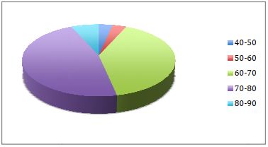
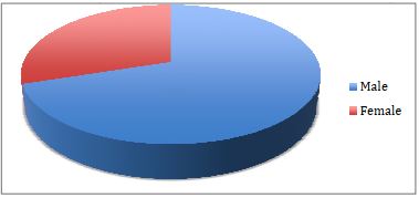
Graph-II: Shows age wise distribution of the subjects

Table I: for Group A for upper limb activity

Pre Intervention RT in sec Post Intervention RT in sec P value Inference
5.52+_1.066 3.73+_0.8433 0.0021 Very significant

Table I states that there is extremely significant in reaction time of upper extremity functional activity with motor practice with auditory cues.

Table-II: Group B for upper limb activity

Pre Intervention RT in sec Post Intervention RT in sec P value Inference
5.6 4.79 0.1013 Not significant

Table II states that there is no significant change in reaction time of upper extremity functional activity with motor practice with visual cues.

Table-III: Group C for upper limb activity

Pre Intervention RT in sec Post Intervention RT in sec P value Inference
5.72 5.55 0.5367 Not significant

Table III states that there is no significant change in reaction time of upper extremity functional activity with motor practice without any cues.

Table IV: Group A for lower limb activity

Pre Intervention RT in sec Post Intervention RT in sec P value Inference
23.89 18.97 0.024 significant

Table IV states that there is extremely significant in reaction time of lower extremity functional activity with motorm practice with auditory cues.

Table-V: Group B for lower limb activity

Pre Intervention RT in sec Post Intervention RT in sec P value Inference
24.3 19.79 0.1601 Not significant

Table V states that there is no significant change in reaction time of lower extremity functional activity with motor practice with visual cues.


Table-VI: Group C for lower limb activity

Pre Intervention RT in sec Post Intervention RT in sec P value Inference
25.82 25.18 0.8499 Not significant

Table VI states that there is no significant change in reaction time of lower extremity functional activity with motor practice without any cues.

bm_36_03.JPG

Discussion

As per the results of the above study auditory cues are effective in reducing bradykinesia. The group with visual cues and the control group did not show any significant improvement. In case of Parkinson’s disease, it’s the basal ganglia which are affected. External cues might facilitate the movement using other brain regions [16]. The premotor cortex is activated with auditory and visual cues. Normally SMA receives inputs from basal ganglia, initiates self-generated movement and perform the previously learned repetitive movements. External cues bypass the basal ganglia and directly activate premotor cortex. Thus focus is shifted to more conscious efforts other than the automaticity. It was found that selection of particular type of cue was dependent on its initial success [17]. This shows that the external cues are important. That could be the reason why the control group did not show any improvement. In this study the visual cues, in the form of video were not effective. This finding is consistent with the previous research which shows that visual and virtual cues were not effective in Parkinson’s patients [21]. In the present study visual cues were virtual meaning were in the form of video clips and hence may be the conscious efforts for the given tasks were affected as patient also had to concentrate on video simultaneously The potential interference is especially highlighting considering that the visuospatioperceptual system must process both the visual cues and the surrounding simultaneously [22,23,24]. Another explanation for the same could be


open loop explanation which was documented in previous study in context to gait cycle. The findings of the association among the walking gait changes and disease profiles are consistent with the cognitive mechanisms underlying the beneficial effects of visual cues. As the disruption of the BG-SMA system increases with the progression of PD, patients become more reliant on the use of vision to move around. The For example, one possible benefit of visual cues is that it may help PD individuals to focus on their task [25-30] possibly by aiding the ability to use cognitive mechanism and holding short-term memory [31,32] or via greater cortical involvement [29,33-37]. This mode avoids unwanted perception of central or peripheral visual flow from the cues as in the case of an open-loop system. In such a system, the cues move independently of the subject’s motion with the objective of assisting or inducing the visual system and locomotor pattern generators [38]. The potential benefits of open loop systems however, are nullified by their obvious drawback. In open loop system person has to face the challenge of challenge of performing the quick activity while simultaneously creating conflicting sensory inputs to the central integrative mechanisms [39]. The capacity of the auditory system to enhance motor performance is used in neurological therapies [40] for rehabilitation purposes [41]. Different  uditory cues (for example, just a metronome tone, a metronome tone embedded into music or just music), are combined with musical parameters (such as rhythm or metre), to emphasize the regular beats in the auditory rhythm. These well-defined sensory cues help regulate timing and pace in walking [41]. These cues may also act as a ‘timer” that helps to regulate the deficient internal timing and rhythm formation processes in PD [42]. One potential method to stimulate the putamen could be music as a provider of an effective rhythmical cue. This type of externally provided cue may be used as a replacement to the ‘internal clock’ to facilitate synchrony of movements [43]. Other studies [44, 45] support the idea that external cues (in particular rhythmical cues) can modulate the activity within the impaired timing system. This may mean that a regular rhythmic pulse stimulates the putamen activity, facilitating movement and providing an input for sequential movements and impaired automaticity. Moreover, this could compensate for the lack of dopaminergic stimulation. Rhythm can be also perceived visually

and through the tactile sense, but the reaction time of the human auditory system is shorter by 20–50 ms, when compared to visual stimuli; [46]. Therefore, rhythm influences the kinetic system (through synchronization and adjustment of muscles to auditory stimuli), and auditory cues [47–49]. All these hypotheses lead to the logical explanation of auditory cues are effective in improving bradykinesia. This is in consensus with previous study [50]. This study was one time study with a small sample size and no long term effects were assessed. Similar study can be conducted on larger sample size with prospective design and long term effects can be assessed.

Reference

1. Victoria A. Goodwin, Suzanne H. Richards, Rod S. Taylor, Adrian H. Taylor, and John L. Campbell. “The effectiveness of exercise interventions for people with Parkinson's disease: a systematic review and metaanalysis”. Mov Disord. 2008; 23(5).

2. Archibald N., Miller N., Rochester L. Neurorehabilitation in Parkinson disease. Handbook of Clinical Neurology. 2013; 110:435–442. doi: 10.1016/ b978-0-444-52901-5.00037-x.

3. Hughes AJ, Daniel SE, Kilford L, Lees AJ. Accuracy of clinical diagnosis of idiopathic Parkinson's disease: a clinico-pathological study of 100 cases. J Neurol Neurosurg Psychiatry. 1992 Mar; 55(3):181-4.

4. Sethi K. Levodopa unresponsive symptoms in Parkinson disease. Mov Disord. 2008; 23 Suppl 3:S521- 33. doi: 10.1002/mds.22049.

5. Albin RL, Young AB, Penney JB. The functional anatomy of basal ganglia disorders. Trends Neurosci. 1989 Oct; 12(10):366-75.

6. DeLong M. R. Primate models of movement disorders of basal ganglia origin. Trends in Neurosciences. 1990; 13(7): 281–285. doi: 10.1016/ 0166-2236 (90)90110-V

7. Obeso JA, Rodríguez-Oroz MC, Rodríguez M, Lanciego JL, Artieda J, Gonzalo N, Olanow CW. Pathophysiology of the basal ganglia in Parkinson's disease. Trends Neurosci.2000 Oct; 23(10 Suppl):S8-19.

8. Benarroch E. E. Intrinsic circuits of the striatum. Complexity and clinical correlations. Neurology.


2016; 86(16):1531–1542. doi: 10.1212/wnl. 0000000000002599.

9. Oorschot D. E. Total number of neurons in the neostriatal, pallidal, subthalamic, and substantia nigral nuclei of the rat basal ganglia: a stereological study using the cavalieri and optical disector methods. Journal of Comparative Neurology. 1996; 366(4): 580–599.

10. Obeso JA, Rodriguez-Oroz MC, Rodriguez M, DeLong MR, Olanow CW. Pathophysiology of levodopa-induced dyskinesias in Parkinson's disease: problems with the current model. Ann Neurol. 2000 Apr; 47(4 Suppl 1):S22-32; discussion S32-4.

11. Berardelli A., Rothwell J. C., Thompson P. D., Hallett M. Pathophysiology of bradykinesia in parkinson's disease. Brain. 2001; 124(11):2131–2146. doi: 10.1093/brain/124.11.2131.

12. Espay AJ, Beaton DE, Morgante F, Gunraj CA, Lang AE, Chen R. Impairments of speed and amplitude of movement in Parkinson's disease: a pilot study. Mov Disord. 2009 May 15; 24(7):1001-8. doi: 10.1002/ mds.22480.

13. Evarts E. V., Teräväinen H., Calne D. B. Reaction time in Parkinson's disease. Brain. 1981; 104(1):167– 186. doi: 10.1093/brain/104.1.167.

14. Jahanshahi M., Brown R. G., Marsden C. D. Simple and choice reaction time and the use of advance information for motor preparation in Parkinson's disease. Brain. 1992; 115(2):539–564. doi: 10.1093/ brain/115.2.539.

15. A. Pascual-Leone, J. Valls-Solé, J. P. Brasil-Neto, L. G. Cohen, and M. Hallett, “Akinesia in Parkinson's disease. I. Shortening of simple reaction time with focal, single-pulse transcranial magnetic stimulation,” Neurology, 44(5). pp. 884–891, 1994.

16. Dibble LE, Hale TF, Marcus RL, Gerber JP, LaStayo PC. High intensity eccentric resistance training decreases bradykinesia and improves Quality Of Life in persons with Parkinson's disease: a preliminary study. Parkinsonism Relat Disord. 2009 Dec; 15(10):752-7. doi:10.1016/j.parkreldis.2009.04.009. Epub 2009 Jun 3.

17. Dietz MA, Goetz CG, Stebbins GT. Evaluation of a modified inverted walking stick as a treatment for parkinsonian freezing episodes. Mov Disord. 1990; 5 (3):243-7.

18. Jahanshahi M, Jenkins IH, Brown RG, MarsdenCD, Passingham RE, Brooks DJ. Self-initiated versus externally triggered movements. I. An investigation using measurement of regional cerebral blood flow with PET and movement-related potentials in normal and Parkinson's disease subjects. Brain. 1995 Aug; 118 (Pt 4): 913-33.

19. Freedland RL, Festa C, Sealy M, Capan A. he effects of pulsed auditory stimulation on various gait measurements in persons with Parkinson's Disease. NeuroRehabilitation 2002; 17(1): 81-7.

20. Suteerawattananon M, Morris GS, Etnvre BR, Jankovic J, Protas EJ. . Effects of visual and auditory cues on gait in individuals with Parkinson's disease. Journal of neurosciences April 2004; 15:219 (1-2): 63-9.

21. Griffin HJ, Greenlaw R, Limousin P, Bhatia K, Quinn NP, Jahanshahi M. The effect of real and virtual visual cues on walking in Parkinson's disease. J Neurol. 2011 Jun; 258(6):991-1000. doi: 10.1007/s00415-010- 5866-z. Epub 2011 Jan 9.

22. Chong RK, Mills B, Dailey L, Lane E, Smith S, Lee KH. Specific interference between a cognitive task and sensory organization for stance balance control in healthy young adults: visuospatial effects. Neuropsychologia. 2010 Jul; 4 8(9):2709-18. doi: 10.1016/j. neuropsychologia.2010.05.018. Epub 2010 May 15.

23. Kemps E, Szmalec A, Vandierendonck A, Crevits L (2005) Visuo-spatial processing in Parkinson's disease: evidence for diminished visuo-spatial sketch pad and central executive resources. Parkinsonism Relat Disord 11: 181-186.

24. Chong RKY, Gibson B, Horton S, Lee A, Mellinger J, et al. (2011) Spatial orientation during eyes closed versus open in the dark: Are they the same? : In review.

25. Devinsky O, Morrell MJ, Vogt BA Contributions of anterior cingulate cortex to behaviour. Brain (1995) 118: 279-306.

26. Kobayashi Y, Inoue Y, Isa T Pedunculo-pontine control of visuallyguided saccades. Prog Brain Res (2004) 143: 439-445.

27. Mathot S, Theeuwes J Visual attention and stability. Philos Trans RSoc Lond B Biol Sci (2011)366: 516-527.


28. Sawamoto N, Honda M, Hanakawa T, Fukuyama H, Shibasaki H Cognitive slowing in Parkinson’s disease: a behavioral evaluation independent of motor slowing. J Neurosci (2002)22: 5198-5203.

29. Sohn YH, Kim GW, Huh K, Kim JS. Dopaminergic influences on the P300 abnormality in Parkinson's disease. J Neurol Sci. 1998 Jun 11; 158(1): 83-7.

30. Steinmetz MA, Constantinidis C. Neurophysiological evidence for a role of posterior parietal cortex in redirecting visual attention. Cereb Cortex 1995; 5: 448-456.

31. Hayes AE, Davidson MC, Keele SW, Rafal RD. Toward a functional analysis of the basal ganglia. J Cogn Neurosci. 1998 Mar; 10(2):178-98.

32. van Schoor NM, Smit JH, Pluijm SM, Jonker C, Lips P. Different cognitive functions in relation to falls among older persons. Immediate memory as an independent risk factor for falls. J Clin Epidemiol. 2002 Sep; 55(9):855-62.

33. Richards M, Cote LJ, Stern Y (1993) The relationship between visuospatial ability and perceptual motor function in Parkinson’s disease. J Neurol, Neurosurg Psychiatry 56: 400-406.

34. Rochester L, Nieuwboer A, Baker K, Hetherington V, Willems AM, et al.(2007) The attentional cost of external rhythmical cues and their impact on gait in Parkinson’s disease: effect of cue modality and task complexity. J Neural Transm 114: 1243- 1248.

35. Sherk H, Fowler GA. Neural analysis of visual information during locomotion. Prog Brain Res. 2001; 134:247-64.

36. Shi LH, Luo F, Woodward DJ, Chang JY. Neural responses in multiple basal ganglia regions during spontaneous and treadmill locomotion tasks in rats. Exp Brain Res. 2004 Aug; 157(3):303-14. Epub 2004 Apr 6.

37. rats. Exp Brain Res 157: 303-314. 69Ferrarin M, Brambilla M, Garavello L, Di Candia A, Pedotti A, et al. (2004) Microprocessor-controlled optical stimulating device to improve the gait of patients with Parkinson's disease. Med Biol Eng Comput 42: 328-332.

38. Closed-Loop VR-Based Interaction to Improve Walking in Parkinson's Disease Chong, J Nov Physiother 2011, 1:1,

39. Thaut M. H., Kenyon G. P., Schauer M. L., McIntosh G. C... The connection between rhythmicity and brain function: implications for therapy of movement disorders. Eng. Med. Biol 1999a. Mag. 18, 101–108 [PubMed]

40. DeBruin,N.,Doan,J.B.,Turnbull,G.,Suchowersky, O., Bonfield, S.,Hu,B.,etal.Walking with music is as a safe and viable tool for gait training in Parkinson’s Disease: the effect of a 13-week feasibility study on single anddual task walking. Parkinson’s disease 9:483530.doi:4061/2010/483530

41. Pastor et al., 1992 M.A. Pastor, J. Artieda, M. Jahanshahi, J.A. Obeso Time estimation and reproduction is abnormal in Parkinson's diseaseBrain: A Journal of Neurology, 1 (115 Pt) (1992), pp. 211-225

42. Journal of Neural Transmission February 2006, Volume 113, Issue 2, pp 175–185204.10. 3109/ 09638288.2013.774060.

43. Benoit CE, Dalla Bella S, Farrugia N, Obrig H, Mainka S, Kotz SA. Musically cued gait-training improves both perceptual and motor timing in Parkinson's disease. Front Hum Neurosci. 2014 Jul 7;8: 494. doi: 10.3389/fnhum.2014.00494. eCollection 2014.

44. Jahanshahi M, Jenkins IH, Brown RG, Marsden CD, Passingham RE, Brooks DJ. Self-initiated versus externally triggered movements. I. An investigation using measurement of regional cerebral blood flow with PET and movement-related potentials in normal and Parkinson's disease subjects. Brain. 1995 Aug; 118 (Pt 4): 913-33.

45. Nombela C, Hughes LE, Owen AM, Grahn JA. Int the groove: can rhythm influence Parkinson’s disease? Neurosci Biobehav Rev (2013) 37:2564– 70.10. 1016/j. neubiorev.2013.08.003.

46. Benjamin WE. A theory of musical meter. Music Percept (1984) 1:355–413.10.2307/40285269.]

47. Jackendoff RS. A Generative Theory of Tonal Music. Cambridge, IN: MIT Press; (1983).

48. Palmer C, Krumhansl CL. Mental representations for musical meter. J Exp Psychol Hum Percept Perform. 1990 Nov; 16(4):728-41.

49. Alfredo Raglio Music Therapy Interventions in Parkinson’s disease: The State-of-the-Art,*Front Neurol. Aug 2015 Published online 2015; 6: 185.